Exercise 9: Nervous Tissue with an Overview of Spinal Cord Anatomy
Dissection Lab: You will need dissection goggles in class
Figure 9.1 One of two cell types found in the nervous system, neurons are responsible for the electrical signals that results in stimulus responses. The image shows a cortical multipolar neuron which constitutes most cells in the brain and spinal cord. This cell was isolated from a mouse and was stained with a fluorescent dye to show the elaborate processes of the neuron.
Exercise 9 Learning Goals
After completing this lab, you should be able to:
- Understand the anatomic and functional divisions of the nervous system
- Examine nervous tissue and identify neurons vs neuroglia cells
- Describe the structure, function, and classification of neurons
- Identify and define structures of the spinal cord though models, images, and specimen dissection
- Be able to describe a reflex arc
- Understand reflexes
 Pre-Lab Activities for Exercise 9
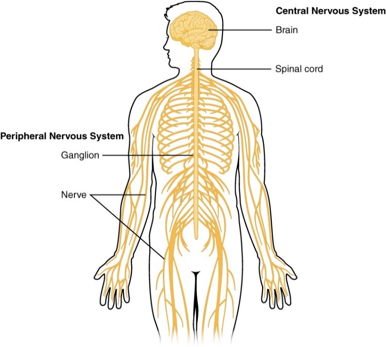
Figure 9.2: Anatomical Divisions of the Nervous System
Pre-Lab Activity 9.1: Divisions of the Nervous System
The nervous system is responsible for perceptions, behaviors, memories, and voluntary movements. It is subdivided based on anatomical or functional description. Anatomically we have a central nervous system which includes the brain and spinal cord and a peripheral nervous system which encompasses the 12 pairs of cranial nerves, 31 pairs of spinal nerves, ganglia, and sensory receptors. The functional division of the nervous system is based on neuron activity including sensory, integrative, and motor functions. Explore the divisions of the human nervous system by completing the table below.
Table 9.1: Divisions of the Nervous System
Division | Structures | Function |
Central Nervous System | ||
Peripheral Nervous System | Somatic: | |
Autonomic: | ||
Enteric: | ||
Sensory | ||
Integrative | ||
Motor |
Figure 9.3: Diversity of neuron types
Pre-Lab Activity 9.2: Introduction to Neurons and Glia
Figure 9.4: Diversity of neuroglia
Nervous tissue is one of the four major tissue types and is composed of two distinct cell types: neurons and neuroglia. Within the two cell types there is further variation based on number of processes, cell body shape and function. Neurons are the electrically excitable cells responsible for sending nerve impulses, while glial cells play a supporting role to neurons; however, glia outnumber neurons approximately 20 to 1.
Study the cells of nervous tissue by completing the tables below.
Table 9.2: Neuron types
Neurons | Function | Shape/structure | Location |
Multipolar | |||
Bipolar | |||
Pseudounipolar | |||
Pyramidal cells | |||
Purkinje cells | |||
Sensory | |||
Motor |
Table 9.3: Glial cell types
Neuroglia | Function | Shape/structure | Location |
Astrocytes | |||
Oligodendrocytes | |||
Microglia | |||
Schwann cells | |||
Ependymal cells |
What is the main difference between the function of neurons and neuroglial cells?
Figure 9.5 Neuron structure
Pre-Lab Activity 9.3: Structure of a Neuron
Neuron structure is unique because these cells are responsible for transmission of electrical signals throughout the nervous system. Neurons are composed of three main parts: dendrites which receive input signals from other cells, cell body and axons which convey output signal to other cells.
Investigate all parts of the neuron by completing the table below.
Table 9.4: Neuron cell parts
Neuron Cell Part | Function |
Cell body | |
Dendrites | |
Axon | |
Axon hillock | |
Synaptic end bulb | |
Myelin sheath | |
Node of Ranvier |
Pre-Lab Activity 9.4: Brain Anatomy
The brain is an extremely important part of the central nervous system protected by the bony skull. It is a jelly-like mass of cells located in the cranium that integrates information, stores memories, makes decisions, receives sensory input, and executes motor output. The mature brain consists of 4 main parts: the brainstem, cerebellum, diencephalon, and cerebrum. The brainstem is further divided into the midbrain, pons and medulla oblongata and the cerebrum is further divided into functional lobes named after cranial bones above that region of the brain (the frontal lobe, temporal lobes, parietal lobes, and occipital lobe). The brain and spinal cord are covered by a 3-layerd non-membrane known as the meninges. Explore the functions of the basic brain structures by completing the table.
Table 9.5: Main parts of the mature brain
Structure | Description | Importance |
Brainstem | Mid brain Pons Medulla oblongata | |
Cerebellum | ||
Diencephalon | ||
Cerebrum | ||
Meninges |
Pre-Lab Activity 9.5: Spinal Cord Anatomy
The spinal cord is an oval structure in the vertebral canal that is continuous inferiorly with the medulla oblongata of the brainstem and connects the central nervous system with peripheral nervous system structures. Explore the functions of important spinal cord structures by completing the table below.
Table 9.5: Structures of the spinal cord
Structure | Description |
Conus Medullaris | |
Filum terminale | |
Cauda equina | |
Central canal | |
Cervical enlargement | |
Lumbar enlargement | |
Meninges | |
Gray Matter | |
White Matter |
Pre-Lab Activity 9.6: Reflexes
The brainstem continues posteriorly as the spinal cord and there are 31 pairs of spinal nerves. The spinal nerves are communication paths between the spinal cord and a specific body region. The spinal cord has internal organization which allows for sensory input and motor output to be processed by the spinal cord. The spinal cord also serves as an integrating center for certain reflexes. A reflex is a fast, involuntary, unplanned sequence of events that occurs in response to a specific stimulus. A reflex is initiated by a nerve impulse stimulated by sensory receptors in the periphery and that impulse is carried to the spinal cord, integrating the information, and responding through the transmission of the nerve impulse to motor neurons and muscle cells in the periphery. There are five components to this reflex circuit, the sensory receptor, sensory neuron, integrating center, motor neuron and effector cell.
Label the five components of a reflex circuit using the diagram.
Figure 9.6: General Reflex Circuit by Ridge Harper
Exercise 9 Activities: Nervous Tissue Activity 9.1: Nervous System Histology
There is a large amount of cellular diversity in the nervous system even though there are only two types of cells: neurons and neuroglia. The diversity originates from differences in cellular morphology, molecular identity, and varying physiology. There are fewer neurons in the nervous system compared to neuroglial cells; however, only the neurons transmit nerve impulses to muscle, glands, and other neurons. The neuroglial cells far outnumber neurons, undergo cell division and are responsible for maintaining the homeostatic environment for the neurons. Some of the neurons you may observe while examining nervous system slides include a multipolar neuron which has many branching processes including many dendrites and an elaborate axon that may or may not be visible. A motor neuron may also be a multipolar neuron; however, it is categorized functionally because its synapses transmit nerve impulses to stimulate muscle fibers. The spinal ganglion slide includes collections of nerve cell bodies as part of the peripheral nervous system. Ganglia are closely associated with spinal nerves that exit on the dorsal side of the spinal cord. Obtain prepared slides of multipolar neurons, motor neurons and spinal ganglia.
A. Examine a multipolar neuron slide:
Using low power magnification identify a multipolar neuron, nuclei of nearby glial cells, dendrites of the neuron, the axon of the neuron and the cell body of the neuron you see.
Label the image after observing the microscope slide: Axon, Cell body (soma), Glial cells, Dendrites
Figure 9.7 Multipolar neurons. Photograph by Gina Profetto
B. Examine a motor neuron slide:
Using low power magnification identify a motor neuron, nuclei of nearby glial cells, dendrites of the neuron, axon of the neuron and cell body of the neuron. Label the image after observing the microscope slide: Glial cell, Dendrites, Axon, Cell Body (Soma)
Figure 9.8 Motor neurons. Photograph by Gina Profetto
C. Examine a spinal ganglion slide:
Using low power magnification identify the cell bodies and nuclei of nearby glial cells in a dorsal root ganglion. Label the image after observing the microscope slide: Glial Cells, Neuron Cell Body, Axons
Figure 9.9 Histology of spinal ganglion slide.
Photograph by Gina Profetto.
Figure 9.10: Transverse section though the spinal cord. Line drawing by Kingsley Dunkley
Activity 9.2: Label the Parts of a Neuron
A neuron is composed of three mains parts including the cell body, which includes the nucleus and carries out the same functions as in other cells, dendrites the many processes originating from the cell body which receives electrical and chemical signals from other neurons and a single axon which may be long or short and conveys electrical signals to the cell synapse communicating with other neurons or muscle cells. Other structures include the axon hillock, node of Ranvier, myelin sheath and synaptic end bulb. Label the parts of the neuron below: cell body (soma), nucleus, dendrites, axon, axon hillock, node of Ranvier, myelin sheath and synaptic end bulb.
Figure 9.11 Unlabeled Neuron
Activity 9.3: Label the Parts of the Spinal Cord
Identify the following structures using the diagram provided: conus medullaris, cervical enlargement, lumbar enlargement, filum terminale and cauda equina.
Figure 9.12 External anatomy of the spinal cord and spinal nerves by Ridge Harper.
Activity 9.4: Spinal Cord Cross Sectional Anatomy
The spinal cord contains both white and gray matter which contains an array of neurons, neuroglia, and neuron processes. Gray matter contains neural cell bodies, with dendrites, and axon terminals. White matter consists of heavily myelinated axons and plays a key role in the neuron’s ability to connect to one another and send sensory ascending and motor descending impulses. Injury to either gray or white matter can interfere with your central nervous system's ability to function.
Label the following areas on the histology image of the cross section: white matter, gray matter, central canal, anterior median fissure, and posterior median sulcus.
Figure 9.13 Histology showing the transverse section of spinal cord.
Label the following areas on the model image of the spinal cord cross section: white matter, gray matter, anterior median fissure, posterior median sulcus, posterior grey horn, grey commissure, anterior grey horn, and central canal.
Figure 9.14 Model of a spinal cord cross sectional segment with anterior and posterior roots attached. Photograph by Gina Profetto
Activity 9.5: Spinal Cord Dissection
The brain and spinal cord are covered by a three-layered protective connective tissue covering known as the meninges. The meninges are composed of the dura mater (outermost layer), the arachnoid mater (middle layer) and the pia mater (innermost layer), attached directly to CNS tissue.
Important Safety Information:
Most of our dissection specimens were preserved in formalin, an aqueous colorless solution that is a suspected carcinogen. In most cases the formalin has been replaced with a nontoxic preservative that may cause mild skin irritation. There is no need for concern provided you follow the guidelines outlined here and wear proper protective equipment.
1) WEAR GLOVES when handling your preserved specimen, tray, tools, or soiled paper towels.
2) WEAR SAFETY GLASSES when actively dissecting.
3) REMOVE GLOVES when you are not working at the lab bench or handling your specimen.
4) ALWAYS CONTAIN YOUR SPECIMEN inside the dissection tray.
5) ALL ORGANIC MATERIAL (identifiable animal parts) should be placed in the orange biohazard bins.
6) IN THE EVENT OF CONTACT: DO NOT PANIC. Wash skin with soap and water; flush eyes with water. There is an eye-wash station at every sink in the lab. Also, please inform your instructor immediately.
Spinal Cord Dissection procedures:
1) Put on safety goggles and gloves
2) Obtain your dissecting tray and instruments
3) Obtain a section of preserved spinal cord
Whole spinal cord:
1. Examine the spinal cord section and observe the connective tissue and fat covering where the spinal nerves exit. The spinal nerves are typically small, white, cylindrical, fibrous structures occurring in pairs about every 1.5 inches along the length of the cord. If the cord were still encased within the vertebral column, one pair of spinal nerves would extend outward between each two vertebrae.
2. Carefully remove the connective tissues until the spinal nerves can be seen. To do this, lift the outermost covering (the dura mater) from the main mass of tissue at one end of the cord. Cut the dura mater along the entire anterior or posterior side of the cord.
4. Try to tear a piece of the dura mater with your fingers. This is a very tough white fibrous connective tissue.
6. The pia mater covers the spinal cord directly, notice that it contains the blood vessels on the surface of the spinal cord.
Cross-section of the spinal cord:
1. Use a sharp scalpel to cut the spinal cord into a transverse section at the point where a pair of spinal nerves exit.
2. Look at the cut end and identify the H-shaped inner core of gray matter. The gray matter is composed of the cell bodies of neurons.
3. The surrounding white matter contains nerve fibers (axons) and appears white due to the myelin which wraps around the axons. The white matter contains bundles of nerve fibers called spinal tracts which conduct impulses to and from the brain.
4. Identify the central canal. Observe the shape of the central canal.
What fills this canal in the living animal?
Clean up procedure:
Dispose of all organic debris in the appropriate biohazard containers and clean the dissecting instruments and tray with soap and water before leaving the laboratory. Do not forget to wash your hands with water and soap, and to disinfect the lab bench.
- What structures make up gray matter?
- What structures make up white matter?
- Why does white matter look white?
- Draw and describe the features observed in a cross section of the spinal cord.
Activity 9.6: Reflex Testing
Consider the various components of the reflex circuit, while testing the following and do not forget about the importance of anatomical position.
- Biceps reflex. With your subject seated on a chair (you should be standing), support their right forearm with your right forearm. Your subject's forearm should be slightly bent at the elbow. Place your thumb over their biceps tendon while applying some pressure to the tendon. Strike your thumbnail with the end of the reflex hammer.
Describe the responses observed.
Repeat the procedure on the left arm. What is the response?
- Patellar reflex. Have your subject sit on the lab bench or a chair so that the legs hang freely. Strike the patellar tendon just inferior to the kneecap with the reflex hammer.
Describe the responses observed.
Test the subject again while they interlock their fingers and pull one hand against the other. Is there a change in the level of activity of the reflex?
- Achilles reflex. Have the subject kneel on a chair and let the foot hang freely over the edge of the chair. Bend one foot to increase the tension on the gastrocnemius muscle. Tap the calcaneal (Achilles) tendon with the reflex hammer.
What is the result?
- Plantar reflex. Stimulate the sole of your subject's foot by moving a blunt object (capped pen or metal end of the reflex hammer) along the sole from the toes to the heel. Describe the movement of the toes.
- In children under two-years old it is normal for the hallux to move upward and outward while the other toes fan out. This is known as the Babinski reflex. Would you expect an adult to exhibit the Babinski reflex? Why or why not?
Post Lab Activity 9.1: Matching
Letter Term | |
_____ Oligodendrocyte | a. These are small nonneural cells that function like phagocytes in the nervous system |
_____ Bipolar cell | b. these cells begin as bipolar cells; however, the dendrite and axon fuse becoming a single process |
_____ Ependymal cell | c. these cells are named after the physiologist that named them and are abundant in the cerebellum |
_____ Microglial cell | d. these cells are found in the CNS lining ventricles of the brain and central canal of the spinal cord |
_____ Motor neuron | e. these cells carry action potentials away from the CNS to muscles and glands in the periphery |
_____ Purkinje cell | f. these neurons are found in the retina and inner ear; they have one main dendrite and one axon |
_____ Pseudounipolar cell | g. these cells are in the CNS and maintain myelin sheaths around axons |
# Term | |
_____ Diencephalon | 1. this structure is superior to the medulla, it is known as a bridge that connects different parts of the brain |
_____ Cerebellum | 2. this area includes an increase in nerves to and from the lower limbs |
_____ Pons | 3. this is the conical structure and the termination of the spinal cord proper |
_____ Meninges | 4. a microscopic channel running the length of the spinal cord and surround by gray commissure |
_____ Lumbar enlargement | 5. a part of the brain that is located medially and includes the hypothalamus and thalamus |
_____ Conus medullaris | 6. the two hemispheres of the forebrain and the largest part of the brain |
_____ Cerebrum | 7. a structure lying posterior to the brainstem and is responsible for balance and coordinating movement |
_____ Central canal | 8. a three-layered membrane covering the brain and spinal cord |
Post Lab Activity 9.2: Label the Spinal Cord Transverse Section
Using the image of a dissected spinal cord segment locate the following areas: anterior fissure, posterior horn, central canal, gray matter, white matter, anterior horn
Figure 9.15 Transverse section of bovine spinal cord taken by ABAC student
A: __________________________
B: __________________________
C: __________________________
D: __________________________
E: __________________________
F: __________________________
Post Lab Activity 9.3: Questions
- Describe the anatomy and location of the spinal cord within the human body.
- How many spinal nerves exit the spinal cord?
- How would you determine dorsal from ventral spinal cord?
- Describe how the spinal cord is suspended in the spinal column.
- What is the overall importance of the spinal cord?
- Myelinated axons are in what area of the spinal cord?
- Would unmyelinated axons and axon cell bodies be in what area of the spinal cord?
- Which type of sensory receptors are you activating to cause reflexes?