Exercise 6: Joints and Muscle Tissue
Figure 6.1 Illustration of skeletal muscles based on anatomical preparations from 17th century scholar Bernardino Genga.
Exercise 6 Learning Goals
After completing this lab, you should be able to:
- Describe the structural and functional classification of joints
- Explain the structure and functions of synovial joints
- Identify major joints by location, classification, and movement
- Communicate the types of movements that occur at synovial joints
- Describe the structural and functional differences between skeletal, cardiac, and smooth muscle
- Explain the anatomical organization of a skeletal muscle including connective tissue, muscle tissue, muscle cells and contractile proteins
- Explain which structures are important for muscle contraction
- Describe the distinct types of fascicle arrangements present in muscle
- Understand how muscles are named
Pre-Lab Activities for Exercise 6Pre-Lab Activity 6.1: Joints
Joints are classified structural as fibrous, cartilaginous, and synovial joints. They are also classified functionally into synarthrosis, amphiarthrosis and diarthrosis. Movements occur at articulation points between bones, specifically synovial joints that are freely movable. The movements that occur at synovial joints include gliding, angular and special movements. Angular movements involve the increase or decrease of an angle between articulating bones and include flexion, extension, lateral flexion, hyperextension, abduction, adduction, and circumduction. Special movements like elevation, depression, protraction, retraction, inversion, eversion dorsiflexion, plantar flexion and opposition occur at specific joints. Use your textbook or notes to complete the following tables.
Term | Definition | Example Joint |
fibrous joint | ||
cartilaginous joint | ||
synovial joint |
Term | Definition | Example Joint |
Synarthrosis | ||
AmphiartHrosis | ||
Diarthrosis |
Pre-Lab Activity 6.2: Skeletal Muscle
Skeletal muscle is organized into bundles of muscle cells. Muscle cells are also known as fibers due to their long, cylindrical appearance. These bundles of muscle fibers are held together by different layers of connective tissues. This connective tissue is continuous with the tendon, which attaches muscle to the bone. There are three layers of continuous connective tissue from superficial to deep. The three layers of connective tissues are:
- Epimysium- covering the entire muscle belly and continuous with tendons
- Perimysium- covering bundles of muscle fibers in units called fascicles
- Endomysium- covering individual muscle fiber/cell
Whole muscles are made up of bundles of fascicles and each fascicle is made up of bundles of muscle fibers.
In the diagram below, fill in the blanks indicating the three layers of muscle connective tissue

C
A
B
Figure 6.2 Organization of muscle (a) muscle belly (b) muscle fascicle (c) muscle fiber.
Pre-Lab Activity 6.3: Myocyte Questions
1. Describe the arrangement of muscle fibers in a skeletal muscle including levels of connective tissues?
2. What is a sarcolemma and what is its main function?
3. Why do muscle fibers require more mitochondria than a typical cell?
4. What is a sarcoplasmic reticulum and what is its main function?
5. Where in a muscle fiber are the contractile units (i.e., sarcomeres) found?
6. What are the two major proteins in a muscle fiber that are responsible for contraction? What other protein plays a significant role in the contraction cycle?
Pre-Lab Activity 6.4: Anatomy of a Muscle Fiber
Use your textbook to fill in the blanks indicating the different organelles of a muscle fiber.
Sarcomere
- Nucleus
- Mitochondrion
- Sarcolemma
- Sarcoplasmic Reticulum
- Myofibril
Figure 6.3 Anatomy of a muscle fiber/cell
Exercise 6 Activities
6.1: Synovial Joints of the Skeleton
Synovial joints are freely movable joints. With bones and skeletal muscle, allow for limb, head, and torso movements. These joints are characterized by having a synovial cavity filled with a viscous lubricating fluid between articulating bones and is enclosed within a two-layered articular capsule. Specific terminology is used to designate the various movements that occur at synovial joints.
Complete the table below.
Movement | Definition | Example Joint |
Gliding | Movement of flat bones in a back and forth or side to side motion | Intercarpal |
Rotation | Movement of a bone around its longitudinal axis | |
Flexion | Elbow | |
Extension | Increase in an angle to normal anatomical position | |
Hyperextension | Atlanto-axial | |
Abduction | ||
Adduction | ||
Circumduction | ||
Elevation | ||
Depression | ||
Inversion | ||
Eversion | ||
Protraction | ||
Retraction | ||
Plantar flexion | ||
Dorsiflexion |
Activity 6:1 Identify Synovial Joint Movements. Your instructor will provide you with images of various movements to complete the activity below.
Image | Describe Movement | Name Joint |
A | ||
B | ||
C | ||
D | ||
E | ||
F | ||
G | ||
H | ||
I | ||
J | ||
K | ||
L |
6.2: Muscle Tissue Histology
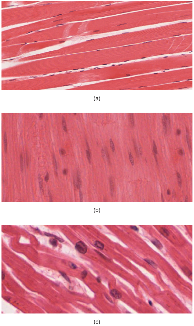 Figure 6.4 Longitudinal sections of the three types of muscle tissue (a) skeletal, (b) smooth, and (c) cardiac.
Activity 6.2: Differences between Skeletal, Cardiac, & Smooth Muscle
Set up your compound microscope and select one muscle composite slide (all three muscle types are present on one slide). When looking at your slide make sure you look at all three types of muscle tissue and identify each one. It is much easier to visualize the features of individual muscle cells in longitudinal orientation. You will need to use your 10X or 40X objectives.
Complete the table below after viewing the types of muscle tissue under the light microscope and use your textbook or lecture notes to find additional information that may not be as easy to see.
Characteristics | Skeletal | Cardiac | Smooth |
Location | |||
Number of nuclei | |||
Location of nuclei | |||
Striated or non-striated | |||
Intercalated discs? | |||
Cell/fiber length | |||
Contraction time |
Within skeletal muscle cells (known as fibers) there are proteins (actin and myosin) that cause the muscle cells to shorten. This shortening will end up moving a bone that the muscle is attached to, and those striations are proteins that help the skeletal muscle to contract. The myofilaments made of proteins are small, but muscles are big!
6.3: Anatomy of the Sarcomere
The sarcomere is the contractile unit of the cell and contains proteins arranged in a way that allows the muscle to contract and shorten (the whole point of a muscle!). In Figure 6.4, bundles of perfectly organized sarcomeres are found within myofibrils (and myofibrils make up each individual muscle fiber).
The contractile proteins within each sarcomere are known as either thick or thin filaments. Thin filaments are made up of the protein actin. Thick filaments are made up of the protein myosin. There are also regulatory proteins troponin and tropomyosin that determine whether thick and thin filaments will interact to initiate a muscle contraction (see steps of sarcomere shortening below)
Figure 6.5 Anatomy of a sarcomere
Activity 6.3: Questions Regarding the Parts of the Sarcomere
- What protein composes thin filaments?
- What protein composes thick filaments?
- Define the different part of a sarcomere
- H zone
- Z lines (discs)
- M line
- I band
- A band
- What is the role of tropomyosin?
- What is the role of troponin?
6.4: Sliding-Filament Theory
Figure 6.6 Visualizing the shortening of a sarcomere using your fingers.
The whole point of a muscle (and sarcomere) is shortened when stimulated. But how does a muscle shorten? And what materials are required to stimulate this shortening?
Interlocking your fingers helps to visualize how a sarcomere can shorten. Place both hands in front of your face with your palms facing you. Your fingers represent the proteins myosin and actin. Your wrists represent Z lines (discs). Interlock your fingers and move them closer together as in Figure 6.6 below. As your fingers (filaments) slide past each other, your wrists (Z lines) get closer together and therefore ‘shorten.’
Steps of Sarcomere Shortening
Sarcomeres shorten by the heads of myosin (thick filament) attaching to myosin binding sites on actin (thin filament). This attachment is called a cross bridge. Using ATP, the myosin head swivels (called the power stroke), bringing the two ends of the sarcomere closer together. Calcium from the sarcoplasmic reticulum is released in response to a muscle action potential from a nerve impulse. Calcium binds to troponin, which causes tropomyosin to change shape and expose myosin binding sites on the actin.
- The myosin head (energized by ATP breaking down to ADP + P) then binds to the actin, forming the cross bridge.
- The myosin head swivels (power stroke), pulling the actin closer to the M line or center of the sarcomere, narrowing the H Zone. ADP +P molecules are released from the myosin head
- The myosin head detaches from the actin when a new molecule of ATP binds to the myosin head
- The new ATP molecule breaks down, releasing energy that re-orientates the myosin head to reposition itself for a new cycle of muscle contraction
Figure 6.7 Steps of one muscle contraction cycle
Activity 6.5: Fascicle Arrangements
Skeletal muscle creates bundles of muscle fibers known as fascicles. The fascicle arrangement within a muscle can aid in its identification. There are five basic fascicle patterns:
- Parallel: Fascicles are arranged side-by-side parallel to each other. The muscle has a strap-like appearance. (Example: Sartorius)
- Fusiform: Fascicles are arranged side-by-side but taper at both ends of the muscle with a bulge in the middle. The muscle takes on a spindle-shaped appearance. (Example: Teres Minor)
- Convergent: Fascicles are broadly distributed at one point of attachment but converge at the other point of attachment. The muscle has a triangular appearance. (Example: Temporalis)
- Circular: The fascicles are arranged in concentric rings around an orifice of the body. The muscle appears circular. (Example: External anal sphincter)
- Pennate: The fascicles are arranged at an oblique angle to a central tendon running through the muscles. If the fascicles are only on one-side of the central tendon, then it is unipennate, and the muscle appears like one-half of a feather. If the fascicles are on both sides on the central tendon, it is bipennate, and the muscle appears like a feather. If the tendon branches several times the fascicles become arranged at multiple angles in curved bundles the muscle is called multipennate.
Figure 6.8: The different types of fascicle arrangements in muscles of the body.
Match the name of the muscle with letters depicted in the image below (Figure 6.9) with fascicle arrangement listed.
Fascicle arrangement | Letter of Muscle Depicted |
Convergent | |
Parallel | |
Bipennate | |
Fusiform | |
Multipennate | |
Circular | |
Unipennate |
Figure 6.9 Skeletal muscle fascicle arrangement and example muscles. Line drawings by Kingsley Dunkley.
Activity 6.6: Anatomy in Clay®
To demonstrate your understanding of origins, insertions, and fascicle arrangement of the muscles, you will use an oil-based clay to form select muscles on the Manikin®. Please follow the instructions below. If you are unsure of what to do, ask your instructor.
- Keep your space clean and organized.
- Keep track of your tools and supplies.
- Feel free to stand or sit when working on your model.
- Move your model around so it is easy to build on but be careful!
- Use dry paper towels to remove clay from hands and tools (DO NOT USE SOAP OR WATER).
- Use red/ terra cotta clay to build muscles.
- Always keep the clay on the green mats.
Clean Up at the end of lab period
1. Take all clay off the model!
2. Roll into balls NO BIGGER than image below and organize into plastic bags.
3. Use dry paper towels to wipe off as much clay as possible from the model and tools used.
4. Return your model to its original place.
Anatomy in Clay®- – Facial Muscles
On your model, place the muscles frontalis and occipitalis, zygomaticus major, orbicularis oculi, orbicularis oris, masseter, and temporalis. Include fascicle orientation.
Muscle | Origin | Insertion | Action |
Frontalis | |||
Occipitalis | |||
Zygomaticus major | |||
Orbicularis oculi | |||
Orbicularis oris | |||
Masseter | |||
Temporalis |
Post-Lab 6 Review
Complete the tables below for axial, upper and lower body joints using information you learned from lab. Use your textbook or notes for reference material.
Upper Body Joints | |||
Joint | Associated Bones/Structures | Movements Possible | |
Acromioclavicular | |||
Glenohumeral | |||
Humeroradial | |||
Proximal radio-ulnar | |||
Distal radio-ulnar | |||
Radiocarpal | |||
Carpometacarpal | |||
Interphalangeal | |||
Spine & Lower Body Joints | |||
Joint | Associated Bones/Structures | Movements Possible | |
Atlanto-occipital | |||
Atlanto-axial | |||
Intervertebral | |||
Sacroiliac | |||
Pubic symphysis | |||
Hip | |||
Patellofemoral | |||
Tibiofemoral | |||
Tibiofibular | |||
Ankle | |||
tarsometatarsal | |||
Interphalangeal | |||
Using the provided image (Figure 6.10) of the head determine the fascicle arrangement for each muscle listed below:
- Sternocleidomastoid (SCM)
Arrangement: ______________
- Orbicularis oris
Arrangement: _______________
- Temporalis
Arrangement: _______________
Figure 6.10: Lateral view of facial muscles.
Matching Terms
_____ Sarcolemma |
|
_____ Sarcoplasmic reticulum |
|
_____ Sarcomere |
|
_____ Perimysium |
|
_____ Epimysium |
|
_____ Endomysium |
|
_____ Actin |
|
_____ Myosin |
Label the parts of the muscle fiber and a sarcomere.
Figure 6.11: Muscle fiber and sarcomere.
Label the parts of the sarcomere and different parts of the thick and thin filaments.
Figure 6.12: Thick and think myofilaments.
Post-Lab Critical Thinking Questions
a) Metatarsophalangeal b) Intertarsal c) Interphalangeal d) Carpometatarsal
- What is the name of the joint between the distal tarsal bones and the metatarsals?
- What is the joint between tarsal bones called?
a) Metatarsophalangeal b) Intertarsal c) Interphalangeal d) Carpometatarsal
- Name the 3 joints that form the knee. Which bones articulate with the patella?
- Name the joint between the metacarpals and the digits?
a) Metacarpalphalangeal b) Hallux c) Interphalangeal d) Carpometacarpal
- Name the joint between the phalanges?
a) Metacarpalphalangeal b) Hallux c) Interphalangeal d) Carpometacarpal
- What is the joint between two carpal bones called?
a) Carpometacarpal b) Intercarpal c) Interphalangeal d) Radiocarpal
11. What is the name of the joint between the distal carpal bones and the metacarpals?
a) Carpometacarpal b) Intercarpal c) Interphalangeal d) Radiocarpal joint
12. List the typical characteristics of synovial joints.