Exercise 4: The Axial Skeleton
Figure 4.1 A basic overview of the bones in the axial skeleton in an adult human
Exercise 4 Learning Goals
After completing this lab, you should be able to:
- Identify bones of the skull
- Distinguish cranial bones from facial bones
- Identify bones of the thoracic cage and vertebral column
- Identify specific bone markings on axial bones
- Understand which bones articulate to facilitate movement
- Understand which markings allow the bones to articulate with one another
Pre-Lab Activity for Exercise 4
Pre-Lab Activity 4.1: Axial Bones
Complete the missing information for each table below. If the box is black, there are no markings to identify for that bone. These are the bones and markings you will be expected to identify on practical exams!
Figure 4.2: Anterior and posterior axial skull
Cranial Bones | Image # | Bone Markings |
Frontal | Supraorbital Foramen | |
Parietal | *Right and left bones | |
External auditory meatus Mastoid process Zygomatic process Styloid process *Right and Left bones | ||
Foramen Magnum | ||
Greater Wings Lesser Wings Optic Canal Foramen Ovale Sella turcica | ||
Cribriform plate | ||
Facial Bones | Image # | Bone Markings |
Nasal | ||
Maxilla (fused) | Infraorbital foramen *Right and Left bones | |
Lacrimal | *Right and Left bones | |
Zygomatic | *Right and Left bones | |
Inferior Nasal Concha | *Right and Left bones | |
Palatine | ||
Vomer | ||
Mandibular condyle (R & L) Mental foramen (R & L) Mandibular notch (R & L) |
Skull Suture | Location |
Between frontal and parietal bones | |
Between parietal and temporal bones | |
Between occipital and parietal bones | |
Between left and right parietal bones |
Vertebrae | # Of bones | Bone Markings to Identify | |
C1 Name: ____________ C2 Name: ____________ | Transverse foramen Anterior and posterior arches (tubercles) Bifid process Dens (odontoid process) | ||
Cervical | Body Spinous process Vertebral foramen Transverse process Inferior articular facets Superior articular Facets Transverse foramen | ||
Thoracic | Body Spinous process Vertebral foramen Transverse process Inferior articular facets Superior articular Facets Superior Costal Facets Transverse Costal Facets | ||
Lumbar | Body Spinous process Vertebral foramen Transverse process Inferior articular facets Superior articular Facets | ||
Sacrum | Sacral promontory Median sacral crest Lateral sacral crests Sacral foramina | ||
Coccyx | Anterior vs posterior |
Thoracic Cage | Region | |
Sternum | ||
Body | ||
Xyphoid process | ||
Rib | True | *Image Color: |
False | *Image Color: | |
Floating | *Image Color: | |
Pre-Lab Activity 4.2: Bone markings
Complete the table below by describing each general bone surface marking.
Bony Landmark | Description |
Foramen | A small or large hole for transmission of artery, vein, nervous tissue |
Sulcus | |
Notch | |
Condyle | |
Facet | |
Process | |
Tuberosity | Roughened area for muscle attachment |
Meatus | Opening |
Trochanter | |
Epicondyle |
Exercise 4: Axial Skeleton
The skull is made up of the brain case and the facial bones. The skull serves to provide protection to soft tissue structures such as the eyeball and the brain. Your study of the skull will consist of first identifying the bone regions, bony landmarks on individual bones, and bones that come together to form the orbit, the nasal cavity, and the cranial fossa.
Activity 4.1: Cranial Bones, Facial Bones, and Suture Joints of the Skull
Identify the cranial bones on a skull model:
- Frontal
- Parietal (R & L)
- Occipital
- Temporal (R & L)
- Sphenoid
- Ethmoid
Identify the facial bones on the skull model:
- Nasal (R & L)
- Maxilla (R & L fused)
- Lacrimal (R & L)
- Zygomatic (R & L)
- Inferior Nasal Concha (R &L)
- Palatine
- Vomer
- Mandible
Identify the sutural joints on a skull model:
Coronal Sagittal Lambdoid Squamous (R & L)
Label the cranial, facial bones and the sutures on the anterior, lateral, posterior and superior views shown below.
Figure 4.6: Skull Model. Photograph by Gina Profetto
Activity 4.2: Orbit
Figure 4.7: Bones of the orbit and associated markings
The orbit is a round bony structure formed by seven skull bones that along with the extraocular muscles hold the eyes in place. The terms below are 3 specific bone markings found on some of the cranial and facial bones.
Using your skull models identify these 3 bones markings:
- Supraorbital Foramen of the Frontal Bone
- Optic Canal of the Sphenoid Bone
- Infraorbital Foramen of the Maxilla bone
Activity 4.3: Sphenoid Bone
The sphenoid bone is the keystone of the cranium and best viewed superiorly looking at the cranial floor. To view this bone, look internally at the skull models. Once you identify the bone, try, and identify the following bone markings.
Lesser Wings of the Sphenoid Bone
- Greater Wings of the Sphenoid Bone
- Foramen Ovale of the Sphenoid
- Sella turcica
Label the bone markings in the view shown below: Lesser wing, Greater wing, Foramen Ovale, Sella turcica
Figure 4.8: Inferior skull. Photograph by Gina ProfettoActivity 4.4: Ethmoid Bone
The ethmoid bone is a delicate bone with a sponge-like appearance and is a major supporting structure of the nasal cavity. To view this bone, look internally at the skull models. Once you identify the bone, try, and identify the listed bone marking.
- Cribriform Plate of the ethmoid bone
Activity 4.5: Temporal Bone
The temporal bone is paired and located inferior-lateral right and left on the skull. Identify the following bone markings on your model.
- Zygomatic Process
- Mastoid Process
- External Auditory Meatus
- Styloid process
Figure 4.9: Lateral skull. Photograph by Gina Profetto.
Activity 4.6: Mandible
The mandible, or lower jaw articulates with the temporal bone via the mandibular condyle and contains the lower teeth. This is the only moveable skull bone. Identify the following bone markings on your model.
- Mandibular (condylar) process (can be located on image above)
- Mental Foramen
- Mandibular Notch (can be located on image above)
Activity 4.7: HyoidThe hyoid bone is a u-shaped bone and does not articulate with any other bone. This unique bone is suspended from the temporal bone by ligaments and muscle, supporting the tongue. Because this bone is near the near tracheal and laryngeal cartilages, manual strangulation is the suspected cause of death when these structures are observed to be damaged or fractured postmortem. Find the hyoid bone on a model and note the greater and lesser horns and the body and label the structures on the diagram below.
Figure 4.10: Hyoid bone and associated markingsThe Thoracic Cage: The thoracic cage is formed by the thoracic vertebrae posteriorly, 12 lateral and anterior ribs right and left, and the anterior medial sternum. The thoracic cage is important for protection of internal organs, including the heart, lungs, great vessels, and liver. The bones of the thorax serve as attachment points for muscles that help facilitate breathing: the diaphragm, and the intercostal muscles. Most of the ribs are attached to the sternum via costal cartilage. Using the whole skeleton and the images below observe how these bones come together to form the thoracic cage.
Activity 4.8: Sternum and Ribs
Locate the following features on the sternum.
- Manubrium
- Body
- Xiphoid Process
Figure 4.11: Sternum and costal cartilage. Photograph by Gina Profetto
View the images below. Note how the head of the rib articulates with the body of the thoracic vertebrae posteriorly. The body of the rib is the curved portion that leads to a flattened rough end, which will attach via coastal cartilages to the sternum. True ribs are ribs that connect directly to the sternum via cartilage. False ribs are ribs that indirectly attach to the sternum, whereas floating ribs only attach to the thoracic vertebrae and do not articulate with the sternum.
Label the images below, note ribs 1-12. Identify the true, false, and floating ribs. Locate the Manubrium, Body, and Xyphoid process of the sternum.
Figure 4.12: Posterior and anterior view of the thoracic cage and associated bones. Photograph by Gina Profetto
The Vertebral Column
The primary function of the vertebral column is protection of the spinal cord. In humans, the vertebral column also gives an upright stance and provides support for the back muscles that help maintain posture. At the base of the vertebral column is the sacrum (part of the pelvic girdle), which supports the bones and muscles of the lower limbs.
The vertebral column begins at the base of the skull, where it articulates with the occipital condyles of the occipital bone. There are 7 vertebrae in the cervical region, 12 in the thoracic region, 5 in the lumbar region, 5 fused sacral vertebra and 3-5 fused coccygeal vertebrae. All vertebrae share some common features.
Activity 4.9: Identify and Label Vertebrae
Figure 4.13: Representative vertebrae. Photograph by Gina Profetto
Label the following features on the image of a vertebrae. These are shared features and will be present on most vertebrae.
- Body
- Spinous process
- Vertebral foramen
- Transverse process (R & L)
- Superior articular Facet (R & L)
- Inferior articular Facet (R & L)
This image has more terms than you need to know, however, it helps visualize how the vertebrae are stacked upon one another connecting via the superior and inferior articular facets. It also demonstrates the intervertebral foramen which only form because the vertebrae are stacked. The spinal nerves exit through these foramina along the length of the vertebral column.
Figure 4.14: Parts of a standard vertebrae and its relationship to other vertebra when stacked in a column
Vertebrae Type | Unique Features |
Cervical | Axis and Atlas; bifurcated spinous process; transverse foramen |
Thoracic | Spinous process long and straight; costal facets for rib articulations found on transverse process and body of rib; resembles face of giraffe |
Lumbar | Shortened spinous process that are elongated in the superior/inferior direction; superior and inferior articular facets are large and more pronounced; resembles face of a moose |
Sacrum | Composed of five fused vertebrae this bone is triangular; the superior forms 2 sacroiliac joint with the hip bones; on the posterior inferior surface is a landmark the sacral hiatus |
Coccyx | Also, triangular shaped, this bone is formed by the fusion of 3-5 vertebrae; in females this bone points inferiorly and in males it points anteriorly |
Cervical Vertebrae:
For atlas (C1) and axis (C2), you must be able to identify the difference between these special bones. These two bones articulate and form a joint called the atlantoaxial joint that allows you to rotate your head side to side.
Identify the superior and inferior sides of the vertebrae, then label the following bone markings for atlas and axis:
Atlas: C1 Axis: C2 Typical Cervical Vertebra
Anterior tubercle Dens (Odontoid Process) Transverse foramina
Posterior tubercle Bifid process
Figure 4.15: Cervical vertebra and associated markings. Photograph by Gina Profetto
For all cervical vertebrae (including atlas and axis) there is an extra marking that allows you to identify that you are looking at a cervical vertebra. On the vertebrae in the image, identify the: transverse foramen of the cervical vertebrae.
Thoracic Vertebrae
The thoracic vertebrae are significantly larger and stronger compared to the cervical vertebrae. First identify the superior and inferior side on the vertebra in the image below and, then identify the:
Figure 4.16: Thoracic vertebrae and associated markings. Photograph by Gina Profetto
- transverse costal facet
- superior costal facet
Lumbar Vertebrae
The lumbar vertebrae are the largest vertebrae with short blunt spinous processes and lack articular surfaces for ribs. Using the image below, identify the shared features and the superior vs. inferior side of the vertebra.
Figure 4.17: Lumbar vertebra and associated markings. Photograph by Gina Profetto
Sacral and Coccygeal Vertebrae
The sacrum and coccyx are first composed of individual vertebrae which fuse between 20-30. The sacrum forms a sturdy foundation along with the hip bones forming the pelvic girdle which supports upper body structures. The coccyx articulates superiorly with the apex of the sacrum.
Figure 4.18: Sacral and Coccygeal vertebra
Practice labeling the following markings: sacral promontory, median and lateral sacral crests, and sacral foramina
Post-Lab 4 Review Questions
1. In what bone(s) would you find the following structures:
- Dens: _______________________________ - Transverse Foramina: ________________
- Lambdoid Suture: _____________________ - Sella Turcica: _______________________
- Cribriform Plate: ______________________ - Zygomatic Arch: _____________________
- External Auditory Meatus: _______________ - Mastoid Process: ____________________
- Mental foramen: ______________________ -Manubrium: ________________________
2. Floating ribs are not truly floating. What bone do they articulate with?
Bone or Structure | Letter |
A | |
B | |
C | |
D | |
E | |
F | |
G | |
H | |
I | |
J | |
K | |
L | |
M | |
N | |
O | |
P | |
Q | |
R | |
S | |
T | |
U |
a) Sternum b) Sphenoid c) Vertebrae d) Hyoid e) Frontal bone
3. Observe the image below and complete the table by identifying the lettered bones and skull structures.
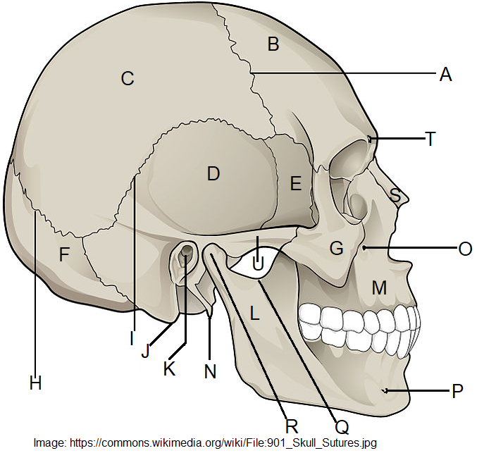 Figure 4.19: Lateral skull
4. The palatine bone is considered a _________ bone.
a) Cranial b) Facial c) Costal d) Sternal e) Appendicular