Exercise 10: Brain Anatomy and Senses
Dissection Lab: You will need dissection goggles in class
Figure 10.1 Colored line engraving by W.H. Lizars of a posterior brain and spinal cord dissection.
Exercise 10 Learning Goals After completing this lab, you should be able to:
- Identify and define structures of the brain though models, images, and specimen dissections
- Describe the structure and function of the major lobes of the brain
- Identify and describe the functions of individual cranial nerves
- Identify the structures of eye and ear
- Understand the differences in somatic touch receptors
- Describe the three main types of color blindness
Pre-Lab Activities for Exercise 10Pre-Lab Activity 10.1: Structure of the Cerebrum
The cerebrum is composed of gray and white matter structures of the right and left hemispheres separated by a deep longitudinal fissure. Each hemisphere is divided into lobes of the brain which have distinct functions and increase the surface area which equates to higher order complex thinking. The increase in surface area is attributed to gyri and sulci: a gyrus is a fold, and a sulcus is a groove between nearby gyri. Gyri and sulci form the four main lobes of the brain. Describe the function and location of the four main lobes of the brain in the table below.
Table 10.1: Lobes of the Brain
Major lobes | Function | Location |
Frontal | ||
Parietal | ||
Temporal | ||
Occipital |
Pre-Lab Activity 10.2: Lateral Structures of the Brain
The lobes of the brain are separated by sulci. In the image below find the central sulcus, Sylvian/lateral sulcus (fissure), parieto-occipital sulcus, frontal lobe, parietal lobe, temporal lobe, occipital lobe, postcentral gyrus, and precentral gyrus.
Figure 10.2: Lobes of the Cerebral Cortex and Major Sulci
Pre-Lab Activity 10.3: Medial Structures of the Brain
The diagram shows a medial view of the human brain. Locate the following structures: frontal lobe, parietal lobe, temporal lobe, occipital lobe, corpus callosum, cerebellum, midbrain, pons, medulla oblongata, thalamus, and pineal gland (body).
Figure 10.3: Medial view of the skull and brain by Ridge Harper.
Table 10.2: Define functions of medial brain structures
Structure | Function |
Midbrain (brainstem) | |
Pons (brainstem) | |
Medulla (brainstem) | |
Corpus callosum | |
Thalamus | |
Hypothalamus |
Pre-Lab Activity 10.4: Cranial Nerves
There are 12 pairs of cranial nerves that originate from the brain inside the cranial cavity and connect the brain to PNS structures. Most of the cranial nerves arise from the midbrain, pons or medulla except for the olfactory nerve which connects directly to the olfactory bulb. Like spinal nerves, the paired cranial nerves are part of the peripheral nervous system and have a name and roman numeral designation. Complete the table below for the cranial nerves.
Table 10.3: Cranial Nerve Function and Their Location
Cranial Nerves | Function | Location |
Olfactory (I) | ||
Optic (II) | ||
Oculomotor (III) | ||
Trochlear (IV) | ||
Trigeminal (V) | ||
Abducens (VI) | ||
Facial (VII) | ||
Vestibulocochlear (VIII) | ||
Glossopharyngeal (IX) | ||
Vagus (X) | ||
Accessory (XI) | ||
Hypoglossal (XII) |
Pre-Lab Activity 10.5: SensesSensation is the conscious or subconscious awareness of the changes internally or externally. Somatic sensations arise from stimulation of sensory receptors embedded in skin or the hypodermis. There are sensory receptors throughout the body, and they are categorized by microscopic structure, receptor location and type of stimulus detected. Special senses are linked to sensory organs that have special receptors and allow us to smell, taste, visualize, hear, and balance. Table 10.4: Sensory Terms
Receptor | Function | Location |
Free nerve endings | ||
Encapsulated nerve endings | ||
Separate cells | ||
Mechanoreceptors | ||
Thermoreceptors | ||
Chemoreceptors | ||
Nociceptors | ||
Photoreceptor | ||
Olfactory receptor cells | ||
Gustatory receptors | ||
Hair cells | ||
Proprioceptors |
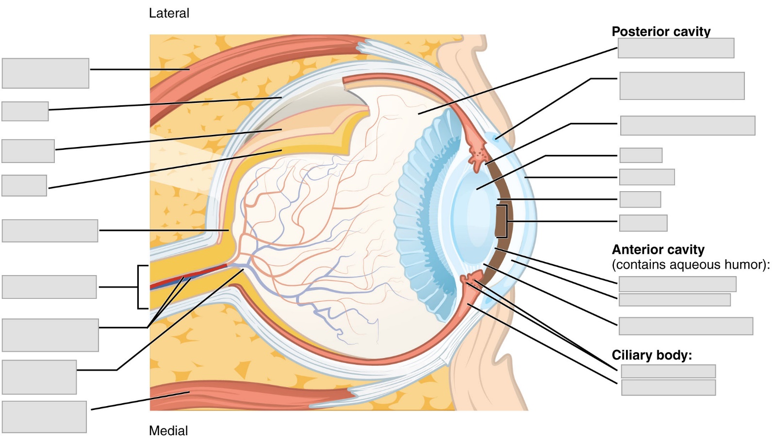
Pre-Lab Activity 10.6: Structure of the Eye
Label the eye using the following terms: lateral rectus, medial rectus, optic disc, sclera, choroid, retina, fovea, optic nerve, retinal veins, and arteries, vitreous, canal of Schlemm, suspensory ligaments, lens, cornea, iris, pupil, posterior chamber, anterior chamber, ciliary process, and ciliary muscle.
Figure 10.4 Label the human eye.
 Pre-Lab Activity 10.7: Structure of the EarLabel the ear using the following terms: eustachian tube, cochlea, round window, cochlear nerve, vestibular nerve, vestibule, stapes, incus, malleus, tympanic cavity, tympanic membrane, auricle, ear canal, external ear, middle ear, and inner ear
Figure 10.5 Label the human ear
Exercise 10 Activities: Brain and Sensory
Lab Activity 10.1: Sheep Brain Dissection
Important Safety Information:
Most of our dissection specimens were preserved in formalin, an aqueous colorless solution that is a suspected carcinogen. Normally, formalin will be replaced with a nontoxic preservative that may cause mild skin irritation. There is no need for concern provided you follow the guidelines outlined here and wear proper protective equipment.
1) WEAR GLOVES when handling your preserved specimen, tray, tools, or soiled paper towels.
2) WEAR SAFETY GLASSES when actively dissecting.
3) REMOVE GLOVES when you are not working at the lab bench or handling your specimen.
4) ALWAYS KEEP YOUR SPECIMEN inside the dissection tray.
5) ALL ORGANIC MATERIAL (identifiable animal parts) should be placed in the orange biohazard bins.
6) IN THE EVENT OF CONTACT: DO NOT PANIC. Wash skin with soap and water; flush eyes with water. There is an eye-wash station at every sink in the lab. Also, please inform your instructor immediately.
1) Put on safety goggles and gloves
2) Obtain your dissecting tray and instruments
3) Obtain a preserved sheep brain
The sheep brain and human brain show many similarities. They both possess a protective membrane called the meninges, which you should observe prior to beginning your dissection. The outermost layer is the dura mater, the toughest layer. The middle layer is known as the arachnoid mater because it resembles a spider web, and the innermost pia mater contains the blood vessels and is attached to the surface of the brain. Using forceps and scissors gently remove the dura. Notice the falx cerebri which separates the right and left hemispheres and the tentorium cerebelli which separates the cerebrum from the cerebellum. When removing the dura mater, falx cerebri and tentorium cerebelli, take care not to remove the brain stem, cerebellum, or optic chiasm.
Identify the following structures on the whole brain or brain model:
Meninges
|
|
Dissection Procedure:
- Gently spread the cerebral hemispheres apart, deep in the longitudinal fissure you will see a thick bundle of white fibers that form the corpus callosum and connects the right and left hemispheres.
- Cut your whole brain along the longitudinal fissure completely through the corpus callosum, midline of the brainstem and cerebellum.
- Separate the hemispheres completely, cutting through the cerebellum and brainstem, then examine the medial structures.
- Below the corpus callosum, you will see a thin membrane, the septum pellucidum and if you pierce this membrane with a sharp tool, you will see part of the lateral ventricle.
- Each hemisphere has one lateral ventricle, c-shaped structures normally filled with cerebrospinal fluid.
- Below the septum pellucidum is a round egg-shaped structure, the thalamus, a relay center for sensory information. There are two thalami, one in each hemisphere connected by a bridge of fibers.
- Gently spread the cerebral hemispheres and the cerebellum (little brain) apart on one half of the dissected brain to reveal the midbrain, seen as two pairs of round swellings (on the whole brain) collectively called the corpora quadrigemina. The larger pair are known as the superior colliculi and the smaller pair are the inferior colliculi. The pineal body is seen directly between the superior colliculi and looks like a little bean and may be cut in half or attached to one half or the other. This structure is important in maintaining a daily sleep-wake cycle.
- The cerebellum is interior and posterior to the cerebrum and connected to the brainstem by three prominent fiber tracts called peduncles. The superior cerebellar peduncle connects the cerebellum with midbrain, the middle cerebellar peduncle connects the cerebellum with the pons and the inferior cerebellar peduncle connects the cerebellum with the medulla. You may carefully remove the cerebellum by cutting through the cerebellar peduncles, revealing the 4th ventricle space.
- The following structures can be located on the ventral surface of the dissected brain. Beneath the frontal lobe of the cerebral hemispheres is where the olfactory bulb is located. The olfactory bulb continues posteriorly as the olfactory tract. Posterior to this tract or anterior to the hypothalamus, the optic nerves undergo a crossing (decussation) known as the optic chiasm.
- Locate the pituitary gland just inferior to the optic chiasm. This gland is connected to the hypothalamus of the diencephalon by a stalk called the infundibulum. The hypothalamus is an important link between the endocrine system and the nervous system.
- The midbrain is inferior to the hypothalamus, the pons is inferior to the midbrain and the medulla oblongata is a posterior extension of the pons. These structures are important for regulating autonomic functions such as consciousness, breathing and blood pressure.
- Identify the frontal lobe, parietal lobe, and occipital lobe, while examining the medial view of the sheep brain.
- Make a coronal section through the cerebral hemisphere just anterior to the thalamus. Examine the section which should reveal gray matter near the surface of the cerebral cortex and white matter beneath this layer. Gray matter represents cell bodies and dendrites of neurons, while white matter is composed of myelinated axons.
- Lastly examine the midsagittal section of the cerebellum. This cut reveals a treelike arrangement of gray matter called folia and white matter called the arbor vitae. The cerebellum is crucial for comparing and coordinating complex muscle movements.
Clean up procedure:
Dispose of all organic debris in the appropriate biohazard containers and clean the dissecting instruments and tray with soap and water before leaving the laboratory. Do not forget to wash your hands with water and soap, and to disinfect the benchtops.
Activity 10.2: Label the Cranial Nerves
Figure 10.6 Label the 12 cranial nerves.
Identify by name and number the 12 pairs of cranial nerves using the diagram provided: Olfactory (I), Optic (II), Oculomotor (III), Trochlear (IV), Trigeminal (V), Abducens (VI), Facial (VII), Vestibulocochlear (VIII), Glossopharyngeal (IX), Vagus (X), Accessory (XI), and Hypoglossal (XII).
Activity 10.3: Somatic Senses
Touch sensations result from stimulation of tactile receptors (mechanoreceptors) in the skin or hypodermis. Crude touch refers to the ability to perceive that something has touched the skin. Fine touch provides more specific information about a touch sensation, such as exactly what point on the body is touched including the shape, size, and texture of the stimulation source. Body areas with few touch receptors are insensitive, whereas those that contain large numbers of touch receptors are more sensitive. This difference can be demonstrated by the two-point discrimination test for touch.
Two-point Discrimination
Two-points using the pointed end of an object (pen or pencil) are applied to the skin and the distance in millimeters between the two points is varied. The subject indicates when they feel two points and when they feel only one.
Procedure:
- Gently, place two pointed objects on the skin on the back of the neck, where there are few receptors. Determine the distance in millimeters when the subject can feel two distinctly different points (start at 1.5mm). Write the distance in the space below.
- Test the back of the neck, fingertip, side of the nose, back of the hand and ankle for each area tested record the distance in millimeters when the subject feels two distinctly different points.
Part of the Body | Distance at which two points can be detected |
Back of neck | |
Tip of finger | |
Side of nose | |
Back of hand | |
Ankle |
Identify Pressure Receptors
Pressure sensation results from stimulation of touch receptors in deeper tissues. Pressure is a sustained sensation that is felt over a larger area compared to touch. It occurs with distortion of deeper tissues. Pressure receptors are found in the skin, mucous membranes, around joints, tendons, muscles, external genitalia, urinary bladder, and pancreas.
- The experimenter touches the skin of the subject (eyes closed) with the point of a piece of colored chalk.
- With eyes still closed, the subject then tries to touch the same spot with a piece of differently colored chalk. The distance between the two points should be measured (mm).
- Repeat using the palm, arm, forearm and back of the neck.
Part of Body | Distance between points touched by chalk |
Arm | |
Palm | |
Forearm | |
Back of neck |
Activity 10.4: Special Senses
Color blindness is an inherited inability to distinguish between certain colors or to detect certain wavelengths of light. It is a sex-linked (X-linked) disorder, which mean that males are more commonly impacted than females. The most familiar form is red-green color blindness, because the genes for those colors are located on the X chromosomes.
Procedure:
Locate the Color Vision Perception Kit and show the subject the 16 shape cards individually to record their response below (NOT the scribble side). Record their results in the table below. If you see all the shapes correctly then you have normal color vision. The reverse side of each shape card demonstrates to normal sighted individuals, what it is like to be color blind.
Card | Actual Shape | Shape Seen by the Subject |
1. | ||
2. | ||
3. | ||
4. | ||
5. | ||
6. | ||
7. | ||
8. | ||
9. | ||
10. | ||
11. | ||
12. | ||
13. | ||
14. | ||
15. | ||
16. |
Is it easy to identify the shapes buried within the scribble card?
Examine the eight photo cards on the normal and then color-blind side. Define deuteranopia, tritanopia and protanopia. In the table below identify the type of colorblindness represented by each card.
Card | Represented form of Color Blindness |
| |
| |
| |
| |
| |
| |
| |
|
Post-Lab 10 Review
Post-Lab Activity 10.1: Brain Anatomy
Below is a medial view of a sheep brain. Circle and label the following structures: brainstem (midbrain, pons, and medulla oblongata), cerebellum, diencephalon (thalamus and hypothalamus), corpus callosum, septum pellucidum, frontal lobe, white matter, gray matter, and optic chiasm.
Figure 10.7: Photograph by Heather Cathcart
Post Lab Activity 10.2: Review Questions
- What is the basic function of?
- Brainstem?
- Cerebellum?
- Diencephalon?
- Cerebrum?
- Which lobes of the brain are separated by the central sulcus? (Circle all that apply)
- Frontal Parietal Insula Occipital Temporal
- Which lobes of the brain are separated by the Sylvian fissure?
- Frontal Parietal Insula Occipital Temporal
- Which parts of the brain are separated by the longitudinal fissure? ________________________________
- Which of the cranial nerves carry special sensory information? ____________________________________
- Which of the cranial nerves carries motor information only? _______________________________________
- Which of the cranial nerves carry both sensory and motor information? _____________________________
- Describe the structures in the pathway of light entering the eye and being processed in the brain.
- Describe the structure in the pathway of sound entering the ear and being processed in the brain.
Post Lab Activity 10.3: Matching
Match the letter to the cranial nerve responsible for the function described. Some letters may be used more than once.
Cranial Nerve | Letter | Function |
Hypoglossal | a. Speech manipulation of food and swallowing | |
Olfactory | b. movement of the head and pectoral girdle | |
Trigeminal | c. hearing and equilibrium | |
Vestibulocochlear | d. decreases heart rate, constriction of respiratory passageways, swallowing, vocalization and coughing, monitors blood pressure and gas levels in blood | |
Glossopharyngeal | e. secretion of saliva and taste from tongue | |
Optic | f. olfaction | |
Oculomotor | g. vision | |
Accessory | h. movement of eyeballs | |
Vagus | j. secretion of tears and saliva, control of facial and middle ear muscles, taste from tongue | |
Facial | k. chewing, touch, pain and heat sensation from scalp, face, and oral cavity | |
Abducens | ||
Trochlear |
Match the cerebral lobe with the correct function and location.
Number | Cerebrum Lobe | Function/Location |
Frontal | 1. Primary somatosensory area of the body; lateral | |
Parietal | 2. auditory association are and learning/memory, inferior | |
Occipital | 3. primary motor area of the body, anterior | |
Temporal | 4. primary visual area, posterior | |
Insula | 5. emotions, autonomic functions |
Match the sensory receptor with the correct definition.
Sensory Receptor | Letter | Location and function |
Free nerve endings | a. these cells are in taste buds and provide information about taste via gustatory microvilli | |
Separate cells | b. these cells are found in the cochlea and in the vestibular apparatus; they transduce sound information and balance information to the brain | |
Proprioceptors | c. these cells are in muscles, tendons, and joints; they provide information about body position | |
Nociceptors | d. these cells are sensitive to stretching and bending of cells and provide information about touch, pressure, and vibration | |
Photoreceptors | e. these are bare dendrites and convey information about pain, tickle, and itch | |
Hair Cells | f. these are specialized cells that synapse with sensory neurons and include hair cells, gustatory receptors, and photoreceptors | |
Mechanoreceptors | g. these cells respond to painful stimuli resulting from physical or chemical damage | |
Gustatory receptors | h. these cells are found in the eye and detect light changes |