Exercise 6: Lymphatic System and
Immune Response
Take Home Lab
Figure 6.1 The above diagram is an anterior view of a dissected human thymus. The thymus is an important lymphatic structure and part of the lymphatic system.
Exercise 6 Learning Goals
At the end of this lab, you will be able to:
- Locate and identify major lymph vessels and organs
- Describe the structure and function of primary and secondary lymphatic organs
- Discuss the cells of the immune system, how they function, and their relationship with the lymphatic system
- Identify the specific cells of the immune system
- Differentiate between innate and adaptive immunity
Pre-Lab Activities for Exercise 6
Pre-lab activity 6.1: Structures of the Lymphatic System
Use your textbook to define the lymphatic terms in the table below.
Term | Definition |
Lymphatic system | |
Lymph | |
Lymphatic capillaries | |
Lymphatic trunks | |
Thoracic duct | |
Cisterna chyli | |
Lymph node | |
Bone marrow | |
Lymphoid nodules | |
MALT (Mucosa Associated Lymphoid Tissue) | |
Spleen | |
Thymus |
Anatomy of the Lymphatic and Immune Systems
The immune system is a complex collection of cells and organs that destroy or neutralize pathogens that would otherwise cause disease or death. The lymphatic system is associated with the immune system to such a degree that the two systems are indistinguishable. The lymphatic system is a subsystem of the circulatory system that consists of a complex network of vessels, tissues, and organs. The lymphatic system carries excess fluids to the bloodstream and filters pathogens from the blood. The swelling of lymph nodes during an infection and the transport of lymphocytes via the lymphatic vessels are two examples of the many connections in this organ system.
Functions of the Lymphatic System
A major function of the lymphatic system is to drain tissue fluids and return them to the bloodstream. Blood pressure causes leakage of fluid from the capillaries, resulting in the accumulation of fluid in the interstitial space. In humans, 20 liters of plasma is released into the interstitial space of the tissues each day due to capillary filtration. Once this filtrate is out of the bloodstream and in the tissue spaces, it is referred to as interstitial fluid. Of this, 17 liters are reabsorbed directly by the blood vessels; however, what happens to the remaining three liters? This is where the lymphatic system comes into play. It drains the excess fluid and empties it back into the bloodstream via a series of vessels, trunks, and ducts. Lymph is the term used to describe interstitial fluid once it has entered the lymphatic system. When the lymphatic system is damaged in some way, such as by cancer cells or destroyed through injury, protein-rich interstitial fluid accumulates in the tissue spaces. This inappropriate accumulation of fluid referred to as lymphedema may lead to serious medical conditions.
Lymphatic capillaries are vessels where interstitial fluid enters the lymphatic system to become lymph fluid. Located in almost every tissue in the body, these vessels are intertwined with the arterioles and venules of the circulatory system. Lymphatic capillaries are formed by a one cell-thick layer of endothelial cells and represent the open end of the system, allowing interstitial fluid to flow into them via overlapping cells. When interstitial fluid pressure is low, the endothelial flaps close to prevent “backflow.” As interstitial fluid pressure increases, the spaces between the cells open, allowing the fluid to enter. Entry of fluid into lymphatic capillaries is also enabled by the collagen filaments that anchor the capillaries to surrounding structures. As interstitial fluid pressure increases, the filaments pull on the endothelial cell flaps, opening them even further to allow easy entry of fluid.
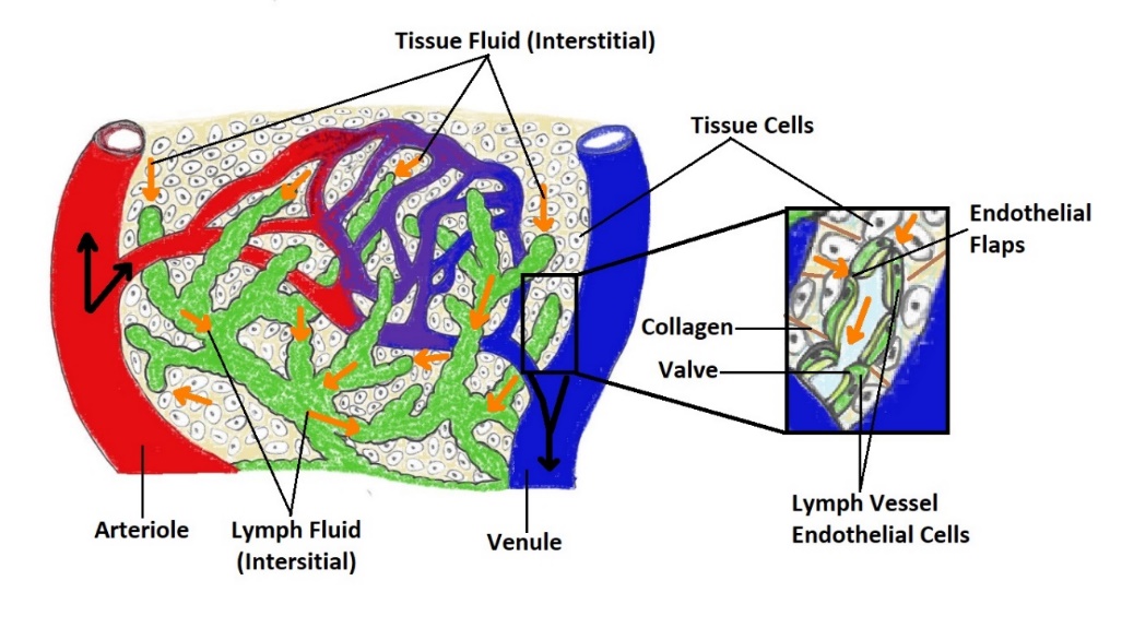 Figure 6.2 Lymphatic capillaries are intertwined with the arterioles and venules of the cardiovascular system. Interstitial fluid slips through spaces between the overlapping endothelial cells that compose the lymphatic capillary (Diagram by Amber Howard).
Figure 6.3 Lymphatic vessels in the arms and legs convey lymph to the larger lymphatic vessels in the torso. The figure depicts the major lymphatic vessels in the arms and legs, with insets that show the detailed anatomy of a lymph node, including the flow of lymph.
Larger Lymphatic Vessels, Trunks, and Ducts
The lymphatic capillaries empty into larger lymphatic vessels, which are like veins in terms of their three-layer structure and the presence of valves. These one-way valves are located close to one another, and each one causes a bulge in the lymphatic vessel, giving the vessels a beaded appearance.
The superficial and deep lymphatics eventually merge to form larger lymphatic vessels known as lymphatic trunks. On the right side of the body, right side of the head, right side of the thorax, and right upper limb drain lymph fluid into the right subclavian vein via the right lymphatic duct. On the left side of the body, the remaining portions drain into the larger thoracic duct, which drains into the left subclavian vein. The thoracic duct itself begins just beneath the diaphragm in the cisterna chyli, a sac-like chamber that receives lymph from the lower abdomen, pelvis, and lower limbs by way of the left and right lumbar trunks and the intestinal trunk.
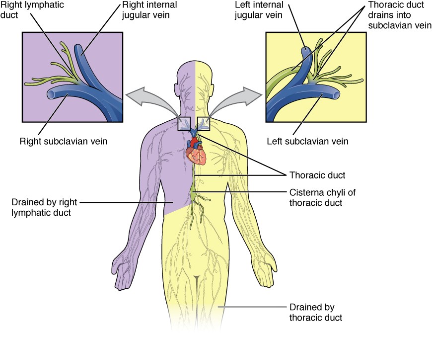 Figure 6.4 The thoracic duct drains a much larger portion of the body than does the right lymphatic duct. The figure depicts the thoracic duct drains the left top of the head, left arm, the left pectoral region, the lower abdomen and legs. The right lymphatic duct drains the right arm, right top of the head, and right pectoral region.
The overall drainage system of the body is asymmetrical. The right lymphatic duct receives lymph from only the upper right side of the body. The lymph from the rest of the body enters the bloodstream through the thoracic duct via all the remaining lymphatic trunks. In general, lymphatic vessels of the superficial lymphatics of the skin follow the same routes as veins, whereas the deep lymphatic vessels of the viscera follow the path of arteries.
Lab Exercise 6: Lymphatic System Anatomy
Activity 6.1: Identify Lymphatic Organs and Associated Structures
Use your textbook to label the lymphatic structures in the diagram.
A. | G. |
B. | H. |
C. | I. |
D. | J. |
E. | K. |
F. | L. |
Figure 6.5 Label Major Lymphatic Structures of the Human Body with letters pointing to specific structures. (Diagram by Gina Profetto)
Activity 6.2: Lymphatic Vessels
Use your textbook to label the major lymphatic trunks, ducts, and blood vessels in the diagram below.
Figure 6.6 Diagram Highlighting Major Lymphatic Vessels and Trunks in the Thoracic Region. (Diagram by Gina Profetto)
A. | E. | I. |
B. | F. | J. |
C. | G. | K. |
D. | H. |
Activity 6.3: Lymphatic Capillaries
Use your textbook to label the structures of the lymphatic capillary.
Figure 6.7 Lymphatic Capillaries and Associated Structures. The figure shows the direction of lymph into capillaries, with boxes to label the individual structures.
Lymphoid Organs
The lymphatic system is commonly divided into the primary lymphoid organs, which are the sites of B and T cell maturation, and the secondary lymphoid organs, in which further differentiation of lymphocytes occurs. Primary lymphoid organs include the thymus, bone marrow, and fetal liver. In humans the thymus and bone marrow are the key players in immune function. All lymphocytes are derived from stem cells in the bone marrow. Stem cells destined to become B lymphocytes remain in the bone marrow as they mature, while prospective T cells migrate to the thymus to undergo further growth. Mature B and T lymphocytes exit the primary lymphoid organs and are transported via the bloodstream to the secondary lymphoid organs, where they become activated by contact with foreign materials that function as antigens.
Secondary Lymphoid Organs and their Roles in Active Immune Responses
Lymphocytes develop and mature in the primary lymphoid organs, but they mount immune responses from the secondary lymphoid organs. A naïve lymphocyte is one that has left the primary organ and entered a secondary lymphoid organ. Naïve lymphocytes are fully functional immunologically but have yet to encounter an antigen to respond to. In addition to circulating in the blood and lymph, lymphocytes concentrate in secondary lymphoid organs, which include the lymph nodes, spleen, and lymphoid nodules.
Lymph nodes function to remove debris and pathogens from the lymph and are thus sometimes referred to as the “filters of the lymph.” Any bacteria in the interstitial fluid are taken up by the lymphatic capillaries and transported to a regional lymph node. Dendritic cells and macrophages within this organ internalize and kill many of the pathogens that pass through, thereby removing them from the body.
Figure 6.8 The thymus lies above the heart. The trabeculae and lobules, including the darkly staining cortex and the lighter staining medulla of each lobule, are clearly visible in the light micrograph of the thymus of a newborn.
Spleen
In addition to the lymph nodes, the spleen is a major secondary lymphoid organ. It is about 12 cm (5 in) long and is attached to the lateral border of the stomach via the gastrosplenic ligament. The spleen is a fragile organ without a strong capsule and is dark red due to its extensive vascularization. The spleen also functions as the location of immune responses to blood-borne pathogens.
Figure 6.9: Lymph nodes are masses of lymphatic tissue located along the larger lymph vessels. The micrograph of the lymph nodes shows a germinal center, which consists of rapidly dividing B cells surrounded by a layer of T cells and other accessory cells.Figure 6.10: (a) The spleen is attached to the stomach. (b) A micrograph of spleen tissue shows the germinal center. The marginal zone is the region between the red pulp and white pulp, which sequesters particulate antigens from the circulation and presents these antigens to lymphocytes in the white pulp.
Activity 6.4: Lymph nodes
Use your textbook to label the structures associated with lymph nodes.
A. | D. |
B. | E. |
C. | F. |
Figure 6.11 Representative Lymph Node and Associate Structures. This figure depicts letters that correspond to specific structures in the lymph node. (Diagram by Gina Profetto)
Lymphoid Nodules
The other lymphoid tissues, the lymphoid nodules, have a simpler architecture than the spleen and lymph nodes in that they consist of a dense cluster of lymphocytes without a surrounding fibrous capsule. These nodules are in the respiratory and digestive tracts, areas routinely exposed to environmental pathogens.
Tonsils are lymphoid nodules located along the inner surface of the pharynx and are important in developing immunity to oral pathogens. The pharyngeal tonsil is at the back of the throat and is sometimes called the adenoid when swollen. Such swelling is an indication of an active immune response to infection. Histologically, tonsils do not contain a complete capsule, and the epithelial layer invaginates deeply into the interior of the tonsil to form tonsils. These structures, which accumulate all sorts of materials taken into the body through eating and breathing, “encourage” pathogens to penetrate deep into the tonsillar tissues where they are acted upon by numerous lymphoid follicles and eliminated.
Mucosa-associated lymphoid tissue (MALT) consists of an aggregate of lymphoid follicles directly associated with mucous membrane epithelia. MALT makes up dome-shaped structures found under the mucosa of the gastrointestinal tract, breast tissue, lungs, and eyes. Peyer’s patches, a type of MALT in the small intestine, are especially important for immune responses against ingested substances. Peyer’s patches contain specialized endothelial cells called M (or microfold) cells that sample material from the intestinal lumen and transport it to nearby follicles so that adaptive immune responses to potential pathogens can be mounted.
Figure 6.12. The pharyngeal tonsil is on the roof of the posterior superior wall of the nasopharynx. The palatine tonsils lay on each side of the pharynx. (b) A micrograph shows the palatine tonsil tissue.
Figure 6.13: Mucosa-associated lymphoid tissue (MALT) of the digestive system- Peyer’s patches.
Bronchus-associated lymphoid tissue (BALT) consists of lymphoid follicular structures with an overlying epithelial layer found along the bifurcations of the bronchi, and between bronchi and arteries. They also have a less-organized structure compared to other lymphoid nodules. These tissues, in addition to the tonsils, are effective against inhaled pathogens.
Activity 6.5: Summary of Lymphatic cells, Tissues, and Organs
Use your required textbook to complete the tables below.
Cell Type | Description/Location | Function |
T helper | ||
T cytotoxic | ||
B cells | ||
Stromal cells |
Mucosa-Associated Lymphatic Tissue
Peyer's patches | ||
Pharyngeal tonsil |
Lymphatic Organs
Lymph nodes | ||
Bone marrow | ||
Spleen | ||
Thymus |
Activity 6.6: Cells of the Immune System
Use your required textbook to label cells A-L in the diagram below.
Figure 6.14 Bood and Lymphatic Cell Lineages. Diagram by Gina Profetto
A. | G. |
B. | H. |
C. | I. |
D. | J. |
E. | K. |
F. | L. |
Organization of Immune Function
The immune system is a collection of barriers, cells, and soluble proteins that interact and communicate with each other in complex ways. The modern model of immune function is organized into three lines of defense based on the timing of their effects. The three timelines consist of the following: the first two lines form the innate immune response while the third the adaptive immune response.
Innate Immune Response
- First line of defense: Barrier defenses such as the skin and mucous membranes, which act immediately to prevent pathogenic invasion into the body tissues
- Second line of defense: The rapid but nonspecific innate immune response, which consists of a variety of specialized cells
Adaptive immune response
- Third line of defense: The slower but more specific and effective adaptive immune response involves many cell types, but is primarily controlled by white blood cells (leukocytes) known as lymphocytes
The cells or formed elements of blood, including those involved in immune response, arise in the bone marrow from hematopoietic stem cells. In contrast with embryonic stem cells, hematopoietic stem cells are present throughout adulthood and allow for the continuous differentiation of blood cells to replace those lost to age or dysfunction. These cells can be divided into three classes based on function:
- Phagocytic cells-which ingest pathogens to destroy them
- Lymphocytes-which specifically coordinate the activities of adaptive immunity
- Granulated cells-which help mediate immune responses against parasites and intracellular pathogens such as viruses
Lymphocytes
As stated above, lymphocytes are the primary cells of adaptive immune responses. The two basic types of lymphocytes, B cells and T cells, are identical morphologically with a large central nucleus surrounded by a thin layer of cytoplasm and both originate from bone marrow. They are distinguished from each other by their surface protein markers and by the molecules they secrete. While B cells mature in red bone marrow, T cells mature in the thymus. Immature T cells migrate from bone marrow to the thymus gland. B cells and T cells are found in many parts of the body, circulating in the bloodstream and lymph, and residing in secondary lymphoid organs, including the spleen and lymph nodes.
B cells are immune cells that function primarily by producing antibodies. An antibody is any of the group of proteins that binds specifically to pathogen-associated molecules known as antigens. An antigen is a chemical structure on the surface of a pathogen that binds to T or B lymphocyte antigen receptors. Once activated by binding to antigen, B cells differentiate into cells that secrete a soluble form of their surface antibodies. These activated B cells are known as plasma cells.
Plasma Cells
A plasma cell is a B cell that has differentiated in response to antigen binding and has thereby gained the ability to secrete soluble antibodies. These cells differ in morphology from standard B and T cells in that they contain a large amount of cytoplasm packed with the protein-synthesizing machinery known as rough endoplasmic reticulum.
T Cells
The T cell does not secrete antibodies but performs many functions in the adaptive immune response. Different T cell types could either secrete soluble factors that communicate with other cells of the adaptive immune response or destroy cells infected with intracellular pathogens.
Natural Killer Cells
A fourth important lymphocyte is the natural killer cell, a participant in the innate immune response. A natural killer cell (NK) is a circulating blood cell that contains cytotoxic (cell-killing) granules in its cytoplasm. It shares this mechanism with the cytotoxic T cells of the adaptive immune response. NK cells are among the body’s first lines of defense against viruses and certain types of cancer.
Activity 6.7: Cells of the Immune System
Use your required textbook to complete the table below.
Cell | Description/Function |
Macrophage | |
Neutrophil | |
Eosinophil | |
Basophil | |
Dendritic | |
Reticular | |
T cell | |
Natural killer | |
Plasma cell |
Activity 6.8: Labeling Lab Models
Using various models in the lab to identify the following lymphatic structures. Images have been provided of these models below in Figure 6.15, 6.16, and 6.17.
|
|
|
|
|
|
|
|
|
|
|
|
|
|
Figure 6.16: Human model of lymphatic structures. Photograph by Amber Howard. |
Figure 6.17: Model of a tibia and fibula articulated with the femur. Photograph by Amber Howard. |
Post-Lab Activity 6.1: Questions
1. What is the function of the thymus?
2. What is the function of the spleen?
3. What are the differences between T cells and B cells?
4. Which of the following cells is phagocytic?
- plasma cell b. macrophage
c. T cell d. natural killer cell
5. Which structure allows lymph from the lower right limb to enter the bloodstream?
- thoracic duct
- right lymphatic duct
- right lymphatic trunk
- left lymphatic trunk
6. Which of the following cells is important in the innate immune response?
- B cells
- T cells
- macrophages
- plasma cells
7. Which of the following cells would be most active in early, antiviral immune responses the first time one is exposed to pathogen?
- macrophage
- T cell
- neutrophil
- natural killer cell
8. Which of the lymph nodules are most likely to see food antigens first?
- tonsils
- Peyer’s patches
- bronchus-associated lymphoid tissue
- mucosa-associated lymphoid tissue
9. Which of the following signs is not a
characteristic of inflammation?
- redness
- pain
- cold
- swelling
10. Which of the following is not important in the antiviral innate immune response?
- interferons
- natural killer cells
- complement
- microphages
11. Enhanced phagocytosis of a cell by the binding of a specific protein is called ________.
- endocytosis
- opsonization
- anaphylaxis
- complement activation
12. Which of the following leads to redness or inflammation?
a. inflammation b. anaphylactic shock
c. obesity d. complement activation
13. What lymph organ is circled in green on the image below?
Figure 6.17: Model of a human abdominopelvic cavity. Photograph by Amber Howard.
Post-Lab Activity 6.2: Matching
Use your textbook to fill in the space with the appropriate letter on the right, these describe the basic functions of the immune system.
Letter | Terms | Function |
Innate Immunity | A. redness, warmth, swelling, and pain because of infection | |
Adaptive Immunity | B. slow and possesses memory of previous antigen | |
Antigen | C. first and second lines of defense of the immune system | |
Cell Mediated Immunity | D. proteins produce by cells to coordinate an immune response | |
Antibody Mediated Immunity | E. foreign substance that induces immune response | |
Surface Barrier | F. over 30 proteins secreted by the liver that help to destroy antigen | |
Phagocytes | G. cells capable of engulfing antigen | |
Cytokines | H. destruction of antigen by B cell | |
Inflammation | I. destruction of intracellular antigen | |
Complement System | J. skin and antimicrobial protein |